|
A 65 year old white female retired RN was admitted to Munson Medical Center for chronic pulmonary infiltrates, cough, and hypoxemia.
Previously healthy, a former smoker (she quit in 1980), she had first become aware of an annoying cough twenty months prior to admission. Initially she believed that it represented a cough related to "post-nasal drip." Eight months prior to admission she and her husband embarked on a four month long round the world trip which would involve visits to China, Thailand, Cambodia, Australia, Easter Island, Egypt and Europe. At about the time of her return from this trip her cough became notably worse and productive, and she first sought medical attention. During an evaluation at a local hospital her CXR showed an large area of consolidation involving her right upper lobe. CT scan showed a dense consolidation likewise in the RUL with smaller infiltrates in the RML and RLL as well as in the lingula of the left upper lobe. She was treated with antibiotics including levofloxacin, ceftriaxone, and azithromycin.
Sputum smears and cultures and legionella and pneumococcal urinary antigens were non-diagnostic at the time of her initial evaluation. However, several weeks later an AFB culture obtained at the time of her initial evaluation was reported positive. She was begun on isoniazid (INH), rifampin, and ethambutol. Subsequently, the acid fast organism was identified as Mycobacterium gordonae. Azithromycin was substituted for INH, and rifampin and ethambutol continued. Additional sputa for AFB were ordered to see if Mycobacterium gordonae might be consistently recovered.
Despite therapy she continued to have a productive cough and continued to experience weight loss (20 pounds cumulatively over the preceding several months). She may have had low grade fevers but did not experience chills or sweats. She became progressively more hypoxemic, and as a consequence, was admitted to the hospital for further evaluation.
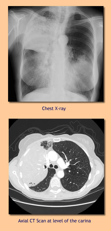
On admission she was afebrile and did not appear to be in distress. Lungs were clear to auscultation. Laboratory evaluation included WBC 6.4, Hgb 14.7. Arterial blood gas (room air) revealed pH 7.45, pCO2 33, pO2 67. CXR and CT scan are pictured at left. In addition to the RUL disease pictured there there also continued to be lesser consolidative changes in the RML, RLL, and lingula of the LUL. Extent of disease appeared to have progressed since prior CT scans. The radiologist favored an inflammatory etiology as there was no volume loss, evident airway compromise, or evident hilar or mediastinal adenopathy.
The patient underwent bronchoscopy with with BAL and transbronchial biopsies. The examining pulmonologist noted thin, non-purulent secretions and essentially normal, patent airways. |